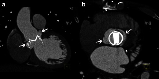
Fig. 7.

Results of MSCT study in case of infective endocarditis on a mechanical aortic valve. MPR reconstructions are shown in the coronal oblique view on the level of the aortic root (a) and on the aortic valve plane view (b). Images show disinsertion of the mechanical prosthetic valve (thin white arrows) and a large pseudoaneurysm around the prosthetic valve (large white arrows)
