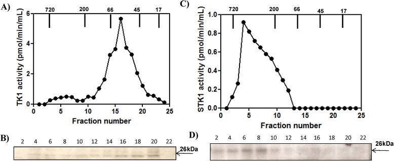
Figure 5.

Acute lymphocytic leukemia (ALL) cell extract and serum analyzed by Superose 12 column chromatography. (A) The thymidine kinase 1 (TK1) activity in the fractions from the ALL cell extract (•). (B) Western blot analyses of the same fractions. (C) Serum TK1 (sTK1) activity in the fractions from the ALL dog (•). (D) Western blot analyses and sTK1 protein in the same fractions. Arrows indicate the elution position of the molecular weight (MW) markers.
