
Figure 6.
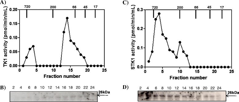
Canine mammary tumor (CMT) (dog No. 23) tumor tissue extract and serum analyzed by Superose 12 column chromatography. (A) Thymidine kinase 1 activity in the fraction from the CMT tissue extract (•). (B) Western blot analyses of the same fractions. (C) Thymidine kinase 1 activity in the fractions from the CMT serum (•). (D) Western blot analyses of the same fractions. Arrows indicate the elution position of the molecular weight (MW) markers. Numbers represent the fast protein liquid chromatography (FPLC) fractions.