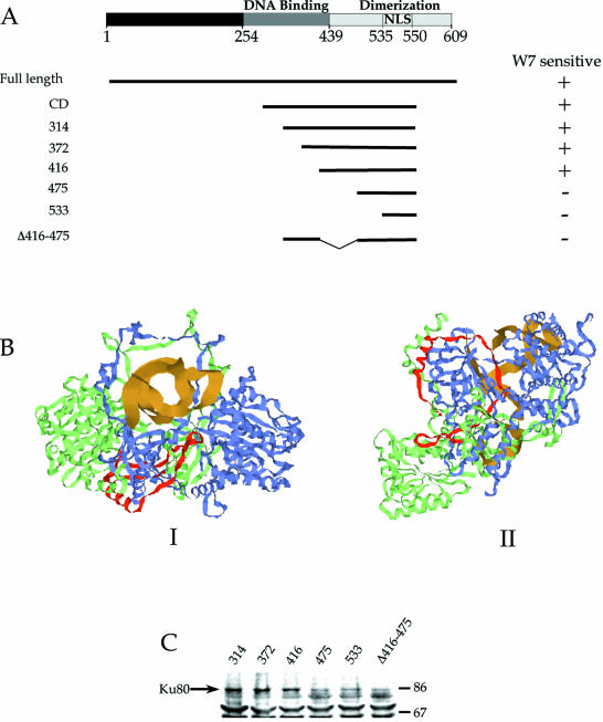
Figure 6.
Mapping of W7 sensitivity to Ku70 functional and structural domains. (A) Loss of sensitivity of protein mobility to W7 corresponds to deletions in the Ku70 dimerization domain. (B) Crystal structure of the Ku70 (green)–Ku80 (blue) heterodimer complexed with DNA (gold). Residues 416–475 of Ku70 are labeled red. I is a view of the complex down the axis of the DNA, and II is turned ∼90° relative to I. (C) Measurement of protein co-immunoprecipitation with Ku80. Each of the Ku70–GFP deletion mutants were expressed and immunoprecipitated using monoclonal antibody to GFP. The samples were separated by protein electrophoresis, and the Ku80 that co-immunoprecipitated with expressed protein was detected by immunoblotting. Molecular weights (in thousands) are indicated on the right.

