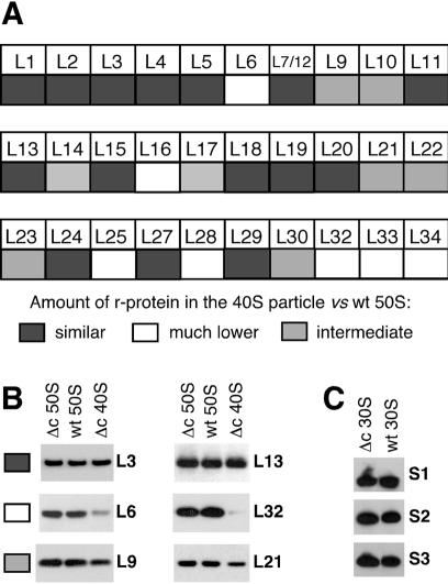
Figure 4.
r-Protein analysis of the 40S and 30S particles from ΔcsdA strain. The ΔcsdA and wt strains were grown at 20°C. r-Proteins were extracted from purified particles and identified and quantified by western blot as described in Materials and Methods. (A) Relative abundance of individual r-proteins in the 40S particle versus the 50S subunit. Grey squares, faint grey squares and white squares correspond to proteins present in similar, lower or much lower amount in the 40S particle, respectively. (B) Western blots illustrating the three classes of proteins defined in (A). Equivalent amounts of 40S particles or 50S subunits (see Materials and Methods) were analysed by western blotting using antibodies against the indicated r-proteins. Δc = ΔcsdA. (C) Western blots showing the presence of r-proteins S1–S3 in the 30S subunits from wt and ΔcsdA strains.

