Figure 3.
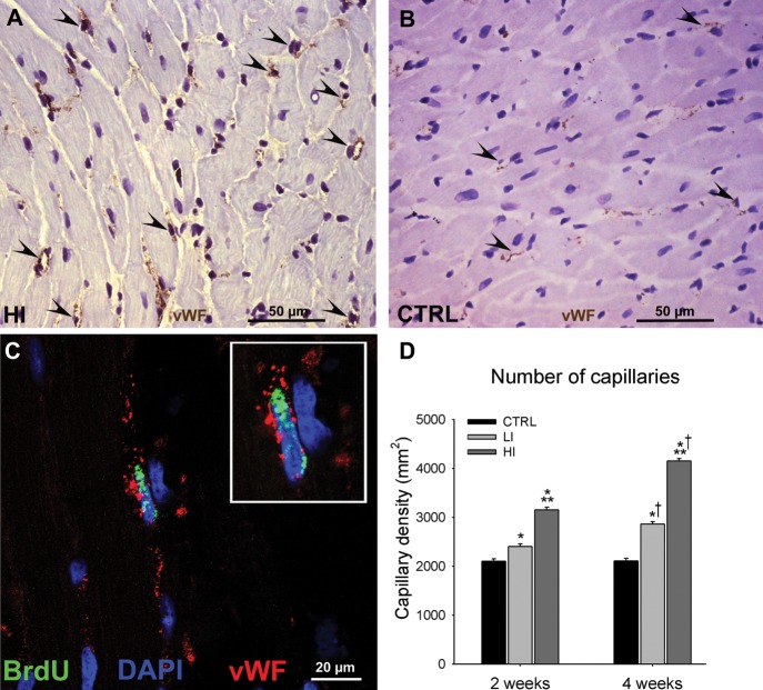
Intensity-controlled treadmill exercise induced angiogenesis. (A and B), Representative images of vWF (brown) capillaries (arrowheads) identified in the left ventricle of a high intensity exercised (A) and CTRL (B) animal. (C) Representative image of a BrdUpos (green) capillary (vWF; red) from the left ventricle of a high intensity exercised animal (Inset is ×2 zoom). (D) The capillary density in the left ventricle of exercising animals was dependent on the exercise intensity and duration (data are mean ± SEM, *P < 0.05 vs. CTRL, **P < 0.05 vs. LI, †P < 0.05 vs. 2 weeks; n = 6 for all).

