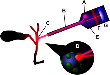
Figure 1.

Principle of fluorescent cholangiography. A source of near-infrared light (A) emits an excitation wave (B), which is directed towards the fluorophore-filled biliary tree at (C). The fluorophore, shown bound to a protein, is excited (D), causing emission of a longer wavelength (E). A filter (F) removes unwanted shorter wavelengths. An image of the fluorescing biliary tree is formed on the charge-coupled device (G), which is then processed for viewing by suitable electronics.
