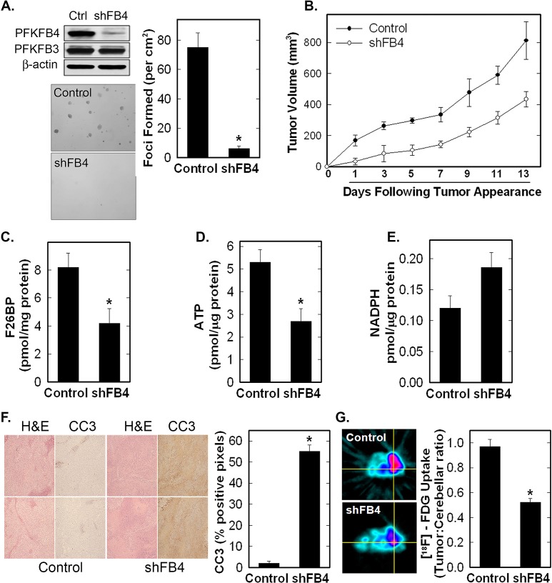
Figure 5. Stable shRNA Knock-Down of PFKFB4 Reduces Tumor Growth, Glucose Uptake and F2,6BP, and Increases Apoptosis.
H460 cells were stably transduced with PFKFB4 (shFB4) or control (Control, Ctrl) shRNA and assessed for PFKFB4 and PFKFB3 protein expression by Western blot analysis (A), soft agar colony formation (A), tumor growth in athymic mice (B). F2,6BP concentration (C), ATP (D), and NADPH (E) were measured in tumors following resection. Data are expressed as the mean ± SD of three experiments. Tumor sections were examined for apoptosis by cleaved caspase 3 (CC3) immunohistochemistry, with representative images on the left and positive pixels quantified on the right (F). The data is depicted as % positive pixels/total pixels ± SD. Tumors in mice were examined for 18F-FDG uptake in vivo by micro-PET imaging. Regions of interest in the tumors and cerebellum were quantified in quadruplicate (right) and representative transverse cuts are shown on the left (G). *p value < 0.001 compared to control shRNA.

