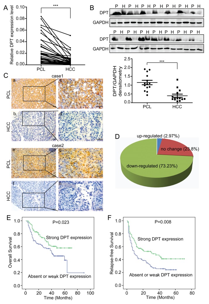
Figure 1. DPT is down-regulated and closely related to patient prognosis in hepatocellular carcinoma (HCC).
(A) Relative mRNA expression of DPT, as determined by qPCR, in 36 pairs of HCC tissues and their paracancerous liver (PCL) tissues. Values are means ± SEM (*** P < 0.001). (B) Western blotting analysis of DPT expression in 14 pairs of HCC and PCL. P represents paracancerous liver tissues and H represents HCC tissues. The densitometric analysis of the results was shown below. Glyceraldehyde-3-phosphate dehydrogenase (GAPDH) was included as a loading control. Values are means ± SEM (*** P < 0.001). (C) Immunohistochemical staining of DPT in HCC and PCL (Original magnification: a, b, e and f, 200x; c, d, g and h, 400x). Scale bars, 10μm. (D) The expression of DPT was down-regulated in 73.23% of the HCC patients. n = 168. (E) Kaplan-Meier analysis of overall survival for the expression of DPT (P = 0.023). (F) Kaplan-Meier analysis of relapse-free survival for the expression of DPT (P = 0.008).

