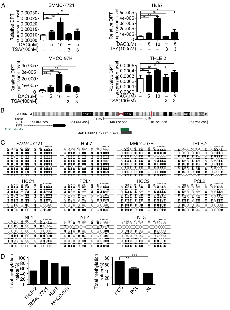
Figure 3. DPT is down-regulated by hypermethylation of DNA promoter.
(A) The reexpression of DPT was evaluated by qPCR in the HCC cell lines and THLE-2 cell line treated with no drug, DAC, TSA or DAC plus TSA. 18s RNA was used as a loading control. Values are means ± SEM (*P < 0.05, **P < 0.01), ns means no significance. (B) Schematic representation of the location of DPT and CpG island within the promoter in chromosome and the primers designed for bisulfite sequencing. (C) Bisulfite-sequencing results of three HCC cell lines (SMMC-7721, Huh7 and MHCC-97H), THLE-2 cell line, 2 pairs of HCC and their paracancerous liver (PCL) tissues and 3 normal liver (NL) tissues. All 13 CpG sites were sequenced. Open circles indicate unmethylated and solid circles represent methylated CpG dinucleotides. (D) The total methylation rates of DPT promoter in the three HCC cell lines (SMMC-7721, Huh7 and MHCC-97H) and THLE-2 cell line, and in HCC, PCL, NL tissues. Values are means ± SEM (**P < 0.01, *** P < 0.001).

