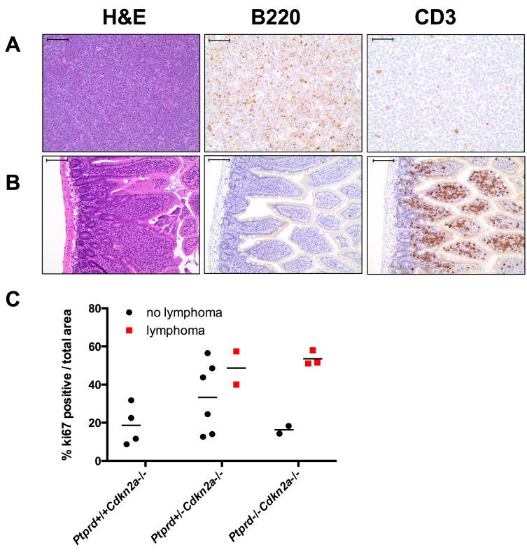
Figure 4. Lymphomas in mice with Ptprd and Cdkn2a loss.
(A) Representative images of Ptprd+/−Cdkn2a−/− B-cell lymphoma in a mesenteric lymph node. Left, scale bar = 100μm; Middle, B220 staining is used to identify B-cells, scale bar = 50μm; Right, CD3 staining is used to identify T-cells, scale bar = 50μm. (B) Representative images of Ptprd−/−Cdkn2a−/− T-cell lymphoma in the small intestine. Left, scale bar = 100μm; Middle, B220 staining, scale bar = 100μm; Right, CD3 staining, scale bar = 100μm. (C) Lymphomas in Ptprd+/−Cdkn2a−/− or Ptprd−/−Cdkn2a−/− mice have similar proliferative indices. Age-matched mesenteric lymph nodes with and without lymphoma were stained with Ki67 by immunohistochemistry.

