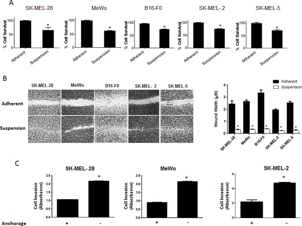
Figure 1. Significant population of melanoma cells resist anoikis in anchorage independent conditions.

(A) SK-MEL-28, MeWo, B16-F0, SK-MEL-2 and SK-MEL-5 cells were cultured under anchorage independent conditions in the plates coated with poly-HEMA for 48 hours and then replated in 24-well plate. The cells were then allowed to attach after which the cell viability was evaluated using Sulforhodamine B assay. The cell survival was compared with the cells cultured under adherent conditions for same time period. Anoikis resistant cells are highly migratory and invasive. (B) Human melanoma cells SK-MEL-28, MeWo, SK-MEL-2, SK-MEL-5 and murine melanoma cells B16-F0 were cultured under adherent or suspension conditions for 48 hours and then replated in a 24-well plate. Confluent monolayers were scratched with 1 mL pipette tip. Wounds were allowed to heal for 16 hours and imaged by microscope. (C) Invasion of SK-MEL-28, MeWo and SK-MEL-2 cells was measured by Boyden's Transwell assay according to the manufacturer's instructions. Values are plotted as mean ± S.D. *, p < 0.05 compared with adherent group. Each experiment was repeated at least three times with similar results.
