
Figure 4. STAT3 deficient cells are sensitive to anoikis and lose migratory potential.
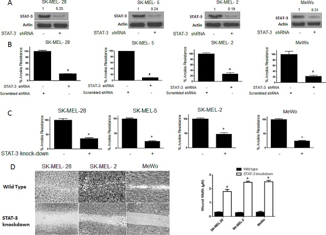
(A-B) SK-MEL-28, SK-MEL-5, SK-MEL-2 and MeWo cells were transfected with STAT3 shRNA for 24 hours after which they were cultured under anchorage independent condition for 48 hours. Cells transfected with scrambled shRNA and cultured under similar conditions were used as control. Percentage of STAT3 silencing was tested by western blotting. Values are plotted as mean ± S.D. *, p < 0.05 compared with control group. Extent of silencing was evaluated by western blotting prior to the experiment and shown by the representative blots for every cell line. (C) STAT3 knock-down human melanoma cell lines SK-MEL-28, SK-MEL-2, SK-MEL5 and MeWo were cultured in anchorage-independent conditions for 48 hours. Cells were replated in a 24-well plate and the viable cells were measured by SRB assay. Wild-type cells of the respective cell lines were used as control. Values are plotted as mean ± S.D. *, p < 0.05 compared with control group. (D) STAT3 knock-down human melanoma cell lines SK-MEL-28, SK-MEL-2 and MeWo cells were cultured in plates coated with poly HEMA. After 48 hours, cells were transferred to a 24-well plate and wound healing assay was performed as described earlier. Respective wild-type cells cultured under similar conditions were used as control. Each experiment was repeated at least three times with similar results