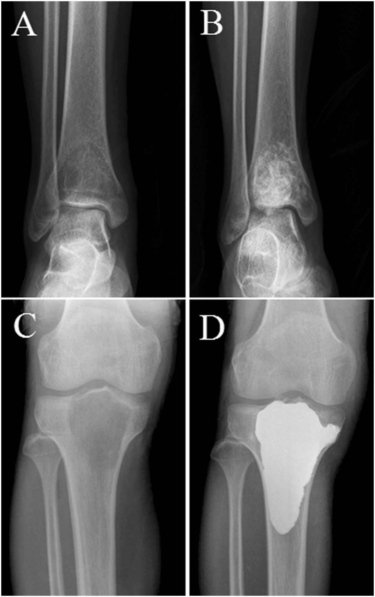
Figure 1.

Typical radiograph of GCT of the long bone. Anteroposterior radiograph shows a lytic lesion in the distal tibia (A) and proximal tibia (C). Anteroposterior radiograph showing the results after agreessive curettage and filling the bone defect with bone grafts (B) and bone cement (D).
