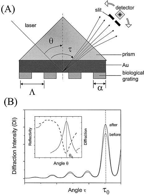
Figure 1.
(A) Schematic geometry for the SPDS. The periodic functional pattern is generated by micro-contact printing (see text for details). θ is the laser incident angle, τ the diffraction angle, α = 42 µm the width of the functional stripes of the surface pattern and Λ = 100 µm the grating constant. (B) Typical diffraction angular scans before and after, for example, hybridization of the target DNA. The inset shows schematically the strong dependence of the monitored diffraction intensity on the surface plasmon coupling angle.

