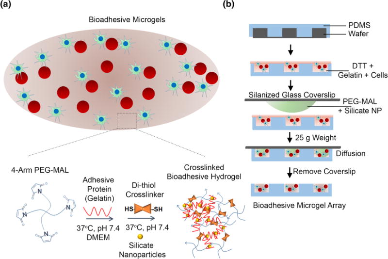
FIGURE 1.

Bio-adhesive, cell encapsulated IPN of PEG-MAL and gelatin-silicate nanoparticles (NP). (a) Schematic of bio-adhesive cell supportive microenvironment consisting of 4-arm PEG-MAL crosslinked with DTT and coated with a stable IPN of gelatin with silicate NP. The 4-Arm PEG-MAL undergoes a Michael-type addition reaction with thiol groups on DTT and gelatin forms an ionic gelation complex with NPs at 37 °C and pH 7.4. The PEG component provides structural support for cells while the gelatin-NP component provides adhesive ligands for cell spreading and signaling. Red spheres represent suspension cells and green cells are anchorage-dependent. (b) Schematic representing microfabrication of bio-adhesive microgels. Component A consisting of a well-mixed solution of gelatin with DTT and media with or without cells and poured onto a PDMS microwell mold. Component B consisting of 4-arm PEG-MAL precursors were mixed with silicate NPs and media was placed on a Sigmacote-coated glass slide and aligned with Component A on each micromold, allowing the polymers to diffuse and mix. After 1 min, glass slides were removed leaving behind an array of cell encapsulated microgels.
