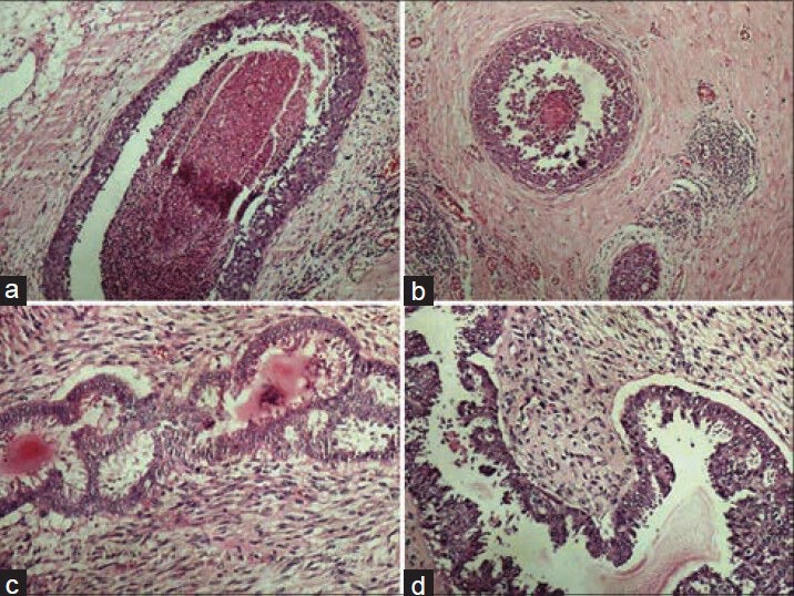
Figure 1.

Photomicrographs showing crisp staining in breast tissue (a) DCIS with conventional (H and E, ×100), (b) DCIS with xylene-alcohol free (XAF) (H and E, ×100), (c) Phyllodes tumor with conventional (H and E, ×200), (b) Phyllodes tumor with XAF (H and E, ×200)
