
Figure 1. MRI.
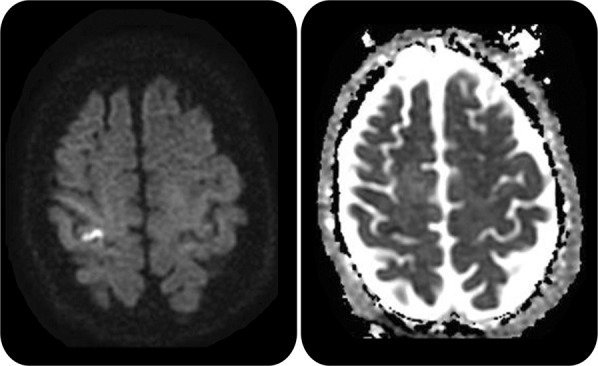
Diffusion-weighted imaging (b1000) (left) shows a hyperintense lesion along the right motor cortex (hand knob) that is hypointense on apparent diffusion coefficient map sequence (right) consistent with an acute infarct.
Official websites use .gov
A
.gov website belongs to an official
government organization in the United States.
Secure .gov websites use HTTPS
A lock (
) or https:// means you've safely
connected to the .gov website. Share sensitive
information only on official, secure websites.
Diffusion-weighted imaging (b1000) (left) shows a hyperintense lesion along the right motor cortex (hand knob) that is hypointense on apparent diffusion coefficient map sequence (right) consistent with an acute infarct.