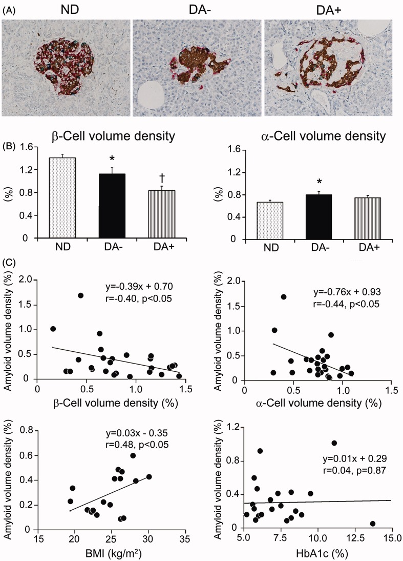
Figure 2.
Islet endocrine cells tetra-immunostained in investigated subjects and correlation of amyloid deposition with β- and α-cell volume density or clinical parameters. (A) Tetra-immunostaining revealed distribution of four types of endocrine cells in the islets of non-diabetic subject (ND), diabetic subject without obvious amyloid deposition (DA−) and with amyloid deposition (DA+) (brown; β-cells, red; α-cells, green; δ-cells, blue; PP-cells). (B) There was a significant reduction of β-cell volume density in amyloid-free diabetic group (DA−) compared to non-diabetic group (ND) (*p < 0.05), and amyloid-rich diabetic group (DA+) showed further reduction (†p < 0.01 versus ND and DA−). α-Cell volume density was greater in DA− compared to ND (*p < 0.05) but that of DA+ was comparable to ND. (C) There was an inverse correlation between β-cell and α-cell volume density with amyloid volume density (p < 0.05). Clinicopathological analysis revealed a correlation between amyloid volume density and BMI (n = 17) (p < 0.05), but not HbA1c (n = 21).

