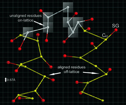
Fig. 2.
Schematic representation of a piece of polypeptide chain in the on- and off-lattice CAS model. Each residue is described by its Cα and side chain center of mass (SG). Whereas Cα values (white) of unaligned residues are confined to the underlying cubic lattice system with a lattice space of 0.87 Å, Cα values (yellow) of aligned residues are excised from templates and traced off-lattice. SG values (red) are always off-lattice and determined by using a two-rotamer approximation (9).

