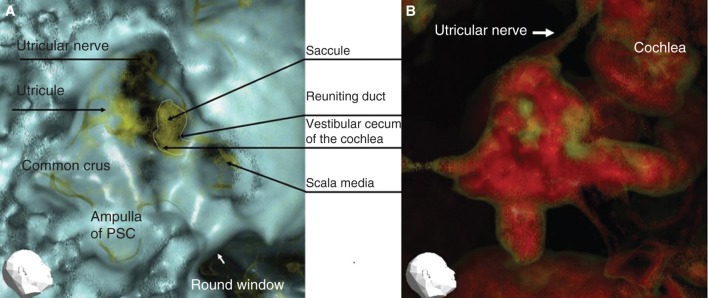
Figure 3.

Representative 3DCT images of the right ear of a patient with Meniere’s disease. (A) Image obtained using CT window values (CTWVs) of CaCO3 (yellow) and bone (blue). (B) Image obtained using CTWV of CaCO3 (yellow) and water (red). PSC, Posterior semicircular canal.
