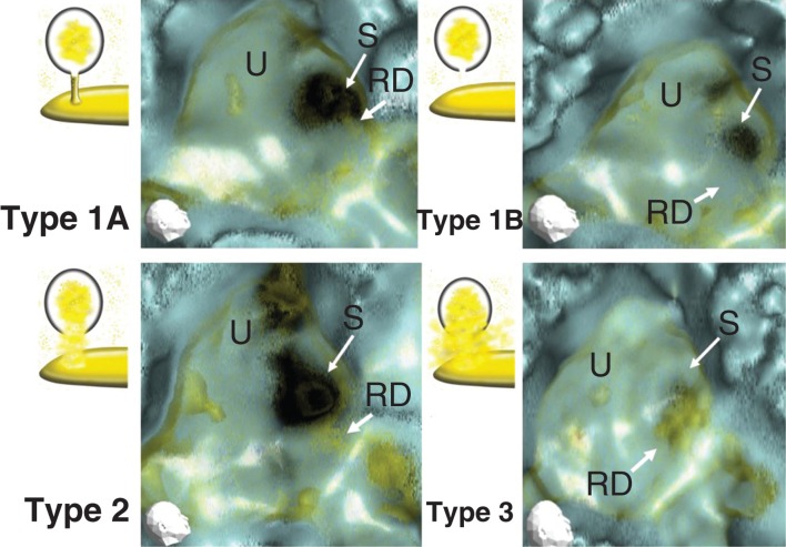
Figure 5.
Representative 3D-CT MICS (membranous image between the vestibular cecum of the cochlea and saccule) classified into three types. Type 1: a neck-like or rod-like narrow line image (type 1A), and no apparent object image in MICS (type 1B). Type 2: irregular and relatively wide line, which however kept the original aspect of the reuniting duct. Type 3: irregular, wide, and condensed MICS with no resemblance to the reuniting duct. RD, reuniting duct; S, saccule; U, utricle.

