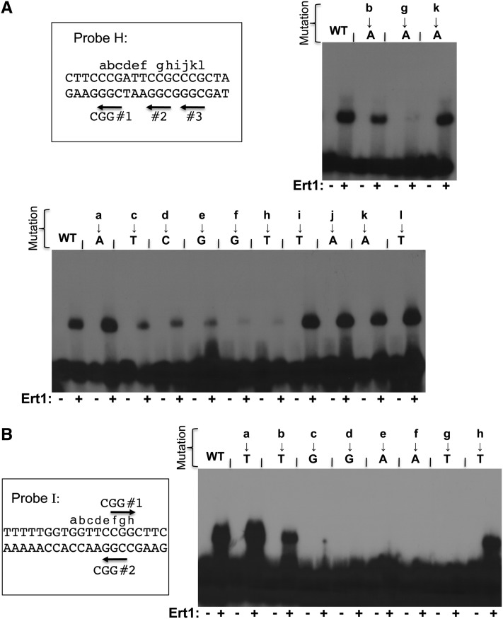
Figure 7.
Ert1 binds to two sites in the PDC1 promoter. (A) Mutational analysis of the binding site “H” of PDC1. DNA sequence of probe H (Figure 6A) is shown at the top as well as the three CGG triplets (labeled CGG nos. 1, 2, and 3). Letters a–l correspond to nucleotides that were mutated. The effect of mutating either CGG triplets to CTG on Ert1 binding was tested by EMSA using the purified DNA binding domain of Ert1 (aa 1–152), as shown in the top right panel. Additional nucleotides were also mutated (bottom panel). WT, wild\x{2010}type. (B) Mutational analysis of the binding site I of Ert1. DNA sequence of probe I (Figure 6A) is shown on the left as well as the two CGG triplets (labeled CGG nos. 1 and 2). Letters a–h correspond to nucleotides that were mutated. Mutants were analyzed by EMSA using the purified DNA binding domain of Ert1 (aa 1–152) (right panel).

