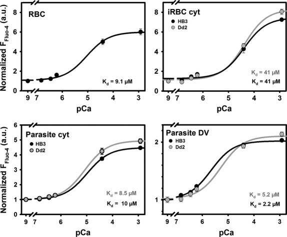
Fig. 4.

Normalized pCa–Fluo-4 steady-state fluorescence relations. In situ calibration relationships were applied to obtain apparent Kd values in the different compartments of HB3- and Dd2-infected erythrocytes. Steady-state Fluo-4 fluorescence intensities are given for different clamped pCa values in ionomycin-permeabilized HB3- or Dd2-infected erythrocytes (iRBC) or non-infected erythrocytes (RBC). Within the parasites, the cytoplasmic and DV compartments were evaluated. Fluorescence values were normalized to the basal fluorescence intensity at pCa 9 (essentially no Ca2+ present) and the apparent Kd values extracted from the sigmoidal relationships. External Fluo-4 AM concentration was 5 μM. The iRBC represents a low-affinity compartment compared to RBC cytosol, most likely due to the drain of Ca2+ into the parasitic compartments. From the reconstructed calibration curves, the apparent Fluo-4 fluorescence derived compartmental Ca2+ concentrations were obtained (see Table 2).
