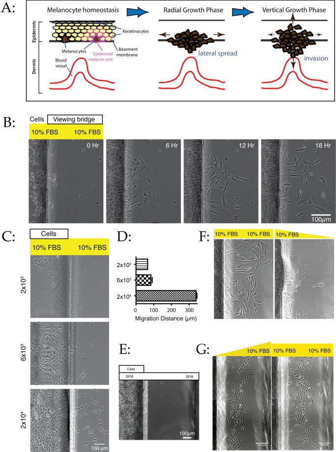
Figure 1. Density-dependent dispersal of melanoma cells.
(A) Schematic showing the stages of melanoma spread. (B) WM239A metastatic melanoma cells dispersing in uniform medium. 2×10⁴ cells were introduced into one reservoir of an Insall chamber containing complete medium with 10% FBS throughout, and observed by time-lapse phase contrast microscopy. See Movie S1. The left side of each image shows the reservoir containing cells, while the right side is the viewing bridge of the chamber. (C–D) Migration is density-dependent. WM1158 metastatic melanoma cells were seeded at different densities in full medium with 10% FBS, and observed as before. At 2×10⁴ cells/well and above, peak migration distances increase sharply, as confirmed by the distance at 17 hours (D; graph shows mean ± SEM). (E) Migration is not driven by production of a repellent. 2×10⁴ WM1158 cells were introduced into a chamber in minimal medium without serum and observed at 17 hours as before. Cells survive and adhere, but do not disperse. (F) Migration is not driven by production of a serum-derived repellent. 2×10⁴ WM1158 cells were introduced into a chamber in minimal medium without serum and observed at 17 hours as before. Cells disperse less efficiently in conditioned medium than in fresh medium. (G) Migration mediated by chemotaxis up a serum gradient is similar to density-induced migration. Left panel: 2×10⁴ WM1158 cells were introduced into a chamber in the presence of a gradient from 0% FBS around the cells to 10% in the opposite reservoir [15]. The cells rapidly migrate towards the well containing serum. Right panel: similar assay with 10% serum in both reservoirs. Panels taken from Movies S3 and S1, respectively.

