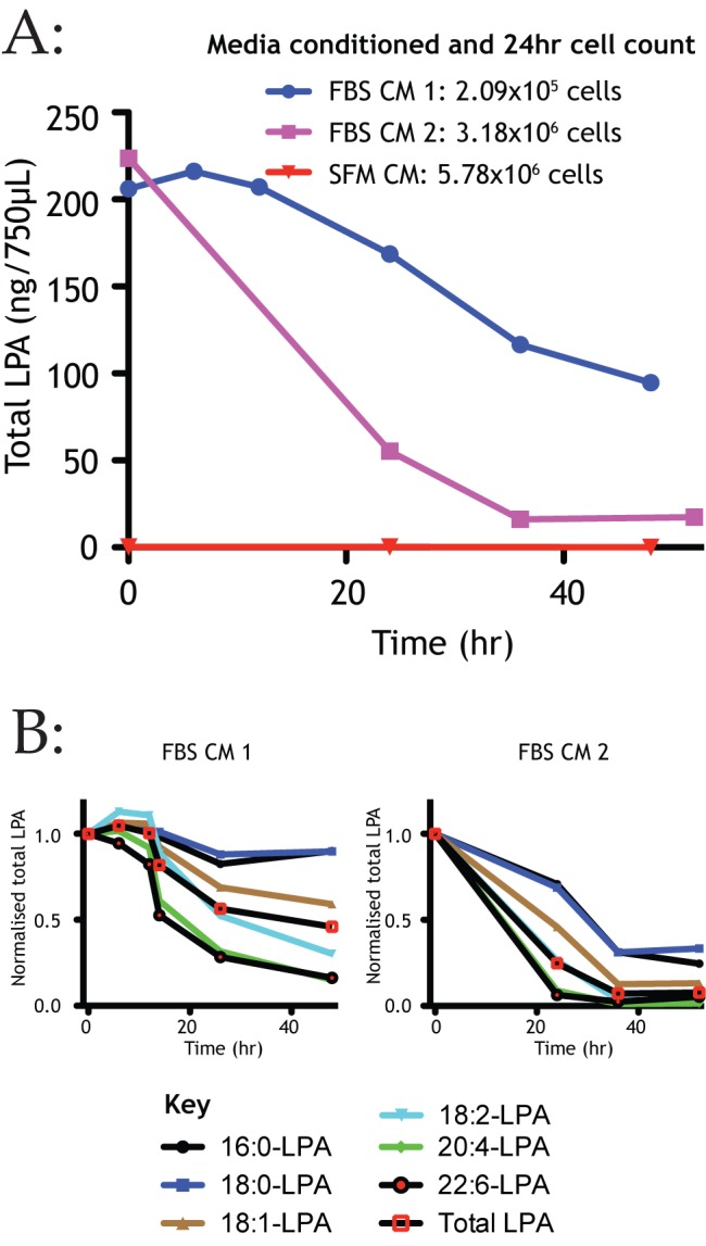
Figure 6. Melanoma cells preferentially break down signalling forms of LPA.

(A) LPA concentration over 48 hours during conditioning of media, both with and without 10% FBS by melanoma cells (WM239A). FBS conditioned media demonstrates density-dependent depletion of LPA as measured by mass spectrometry. LPA remained negligible throughout 48 hours of serum-free conditioning by the same cells. Representative graph. (B) Analysis of LPA subspecies during melanoma cell conditioning demonstrates bioactive isoforms were depleted more rapidly by melanoma cells in both samples. Two representative graphs are shown to illustrate quantitative variability but qualitative consistency.
