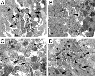
Fig. 5.
High-power light microscopic analysis of spermatids in high-percentage Klhl10 chimeric testes. (A) A tubule contains step 1 spermatids (arrowheads), step 9 (arrows), and steps 12 and 13 spermatids (circled). Note that multiple nuclei of step 12 and 13 spermatids appear to share a common cytoplasm, and several large or small, darkly stained, vacuolated cytoplasmic/cellular bodies (stars) are present within the epithelium. Spermatids appear to slough toward the lumen. (B) A tubule contains steps 8 and 9 spermatids. Two step 8 elongating spermatid nuclei (arrow) share a common acrosome and cytoplasm. (C) Multinucleated step 8 and 9 spermatids (arrows) are sloughing into the lumen, and large vacuolated cellular bodies are present in the seminiferous epithelium. (D) Disorientated and asynchronized spermatid maturation in the seminiferous epithelium. Steps 9 and 10 spermatids (arrows) coexist with step 11 and 12 spermatids (arrowheads) and step 6 and 7 round spermatids (circled). Degenerating cellular bodies (stars) are also present in the epithelium. Lu, lumen. (magnification, ×1,000).

