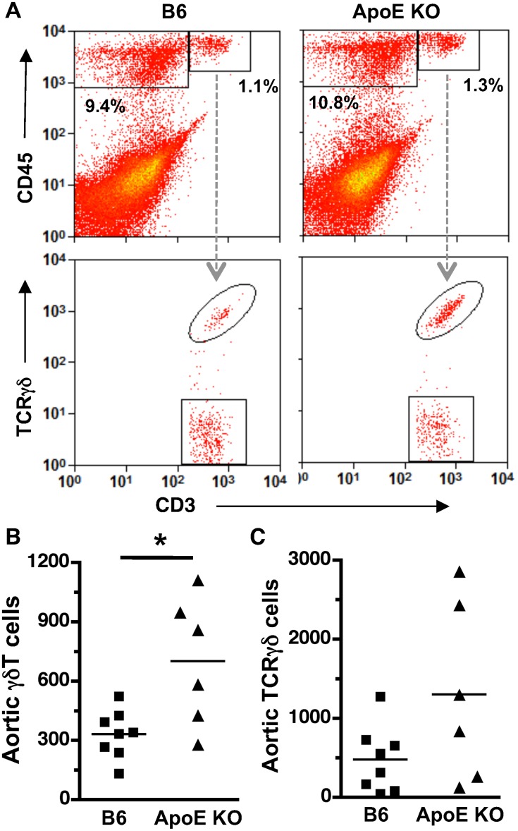
Figure 1. Increased γδT cells in proximal aorta of ApoE KO vs. B6 mice.
Single-cell suspensions from enzyme-digested aortas of 22 wk-old chow-fed mice were stained with anti-CD3-FITC, anti-CD45-PE and anti-TCRγδ-APC and analyzed by FACS. A: Gating strategy and representative FACS plots. B: Bars indicate means; symbols indicate the absolute number of aortic T cells in individual mice. γδT cells (CD3+TCRγδ+) were significantly increased in ApoE KO vs. B6 aorta (*p<0.04). The absolute numbers of conventional αβT cells (CD3+TCRγδ−) were highly variable in individual aortas from ApoE KO mice and were not significantly different from the numbers in B6 mice.

