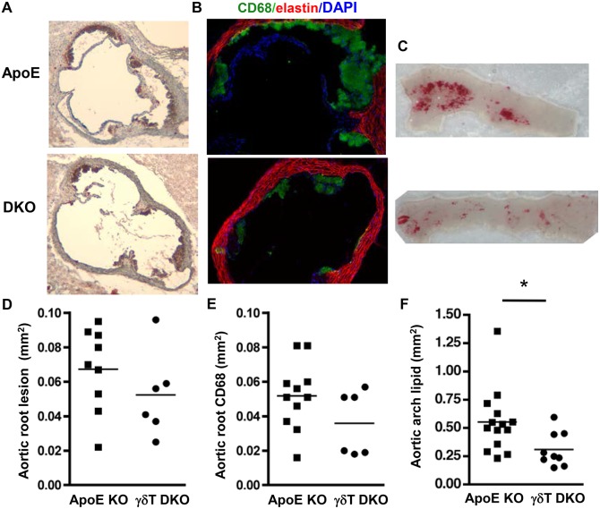
Figure 5. γδT cell deficiency reduces aortic arch lesion area in Western diet-fed ApoE KO mice.
Mice were fed Western diet for 4 weeks and aortic root cross sections were stained with A: ORO and hematoxylin, or B: anti-CD68-Alexa Fluor 488, AlexaFluor 633 to visualize elastin, and DAPI. C: Aortic arch segments were stained with ORO en face. D&E: Charts show total and macrophage-rich lesion areas in the aortic root. The aortic roots of 3 ApoE KO mice and 3 ApoE/γδT DKO mice were excluded from the analysis based on the lack of 3 complete cusps. F: Aortic arch lesions were significantly reduced in ApoE/γδT DKO mice (*p<0.03).

