
FIGURE 3:
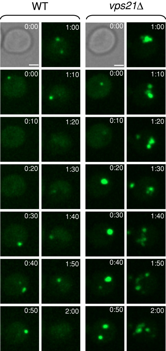
Accumulation of multiple autophagosomes in vps21∆ mutant cells. The dynamics of autophagosomes marked with GFP-Atg8 was determined in wild-type and vps21∆ mutant cells using time-lapse live microscopy. Sequential frames of GFP-Atg8 dots (in 10-min intervals) are shown, with differential interference contrast at time 0. WT and vps21∆ mutant cells were grown in YPD medium until log phase and shifted to SD-N medium for 5 min before the cells were sampled on 2% agar in SD-N for observation by time-lapse video microscopy. Bar, 2 μm. See videos of GFP-Atg8 in WT (Supplemental Movie S1) and GFP-Atg8 in vps21∆ mutant cells (Supplemental Movie S2) for 20-s intervals, playing at 12 frames/s (fps). Results represent three independent experiments.