
FIGURE 6. Signals responsible for lysosomal localization of TRPML1 and TRPML2.
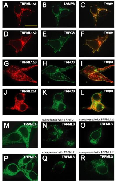
A and B, confocal images of HEK293 cells previously co-transfected with TRPML1Δ1-HA and LAMP3-YFP in which immunofluorescence was performed with anti-HA primary antibodies and Alexa568-conjugated secondary antibodies and the cells were viewed at the indicated excitation wavelengths: 568 nm, TRPML1Δ1-HA, red (A); 488 nm, LAMP3-YFP, green (B). C, merge of A and B. D–L, same as A–C but in HEK293 cells co-transfected with either TRPML1Δ2-HA and TRPC6-YFP, TRPML1Δ3-HA and TRPC6-YFP, or TRPML2Δ1-HA and TRPC6-YFP, respectively. M–O, confocal images of HEK293 cells transfected with vectors encoding the following and viewed at an excitation wavelength of 488 nm: TRPML3-YFP alone (M), TRPML3-YFP and TRPML1 (N), TRPML3-YFP and TRPML1Δ2 (O). P–R, same as M–O except in HEK293 cells transfected with: TRPML3-YFP alone (P), TRPML3-YFP and TRPML2 (Q), or TRPML3-YFP and TRPML2Δ1 (R). The scale bar represents 20 μm.