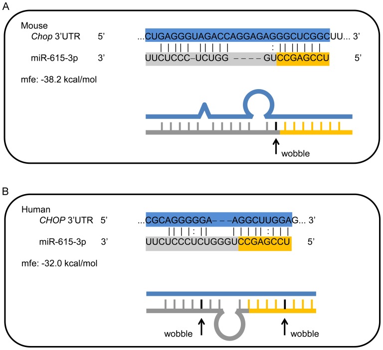
Figure 1. Computationally identified miR-615-3p binding site in Chop mRNA.
(A) Schematic representation of the sequence for mouse Chop mRNA showing a section of the 3′ untranslated region (UTR) and the mmu-miR-615-3p sequence below. The 7-mer binding site is depicted by the solid lines at the 3′ end of the Chop mRNA. (B) Sequence of a section of the human 3′UTR of the CHOP mRNA, with the potential mir-615-3p binding site is depicted. For each binding site, the sequence alignment is shown as well as a schematic line diagram of the predicted complementary regions and bulges. The predicted minimum free energy (mfe) is depicted. Preferences were set to allow G:U wobble bases within the alignment; these are indicated in the figure.

