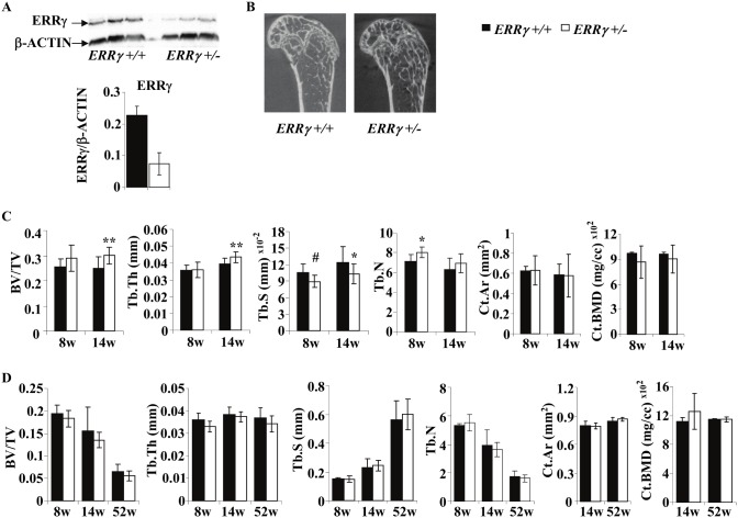
Figure 2. Trabecular bone formation is increased in 14-week old male distal femurs.
(A) Quantification of Western blots revealed that ERRγ protein expression was reduced in whole cell lysates of trabecular bones of 14-week old ERRγ +/− mice compared to ERRγ +/+ mice. (B) Representative µCT images of 14-week old ERRγ +/+ and ERRγ +/− male distal femurs. (C) Quantitative analysis revealed a significant increase in trabecular bone volume fraction (BV/TV) and thickness (Tb.Th), and a decrease in separation (Tb.S) at 14 weeks. Trabecular number (Tb.N) was significantly increased at 8 weeks, but not significantly at 14 weeks (upper panels). There were no significant differences in cortical bone area (Ct.Ar) or bone mineral density (BMD) (lower panel). (D) Analysis on 8, 14 and 52-week old female mice revealed no differences in any bone parameters assessed. Values are expressed as mean ± SD (Male: 8w: N = 7 ERRγ +/+; N = 5 ERRγ +/−; 14w: N = 10 ERRγ +/+; N = 14 ERRγ +/−; Female: 8w: N = 3 ERRγ +/+; N = 5 ERRγ +/−; 14w: N = 10 ERRγ +/+; N = 7 ERRγ +/−; 52w: N = 6 ERRγ +/+; N = 5 ERRγ +/−) * = p<0.05; ** = p<0.01; # = 0.069.

