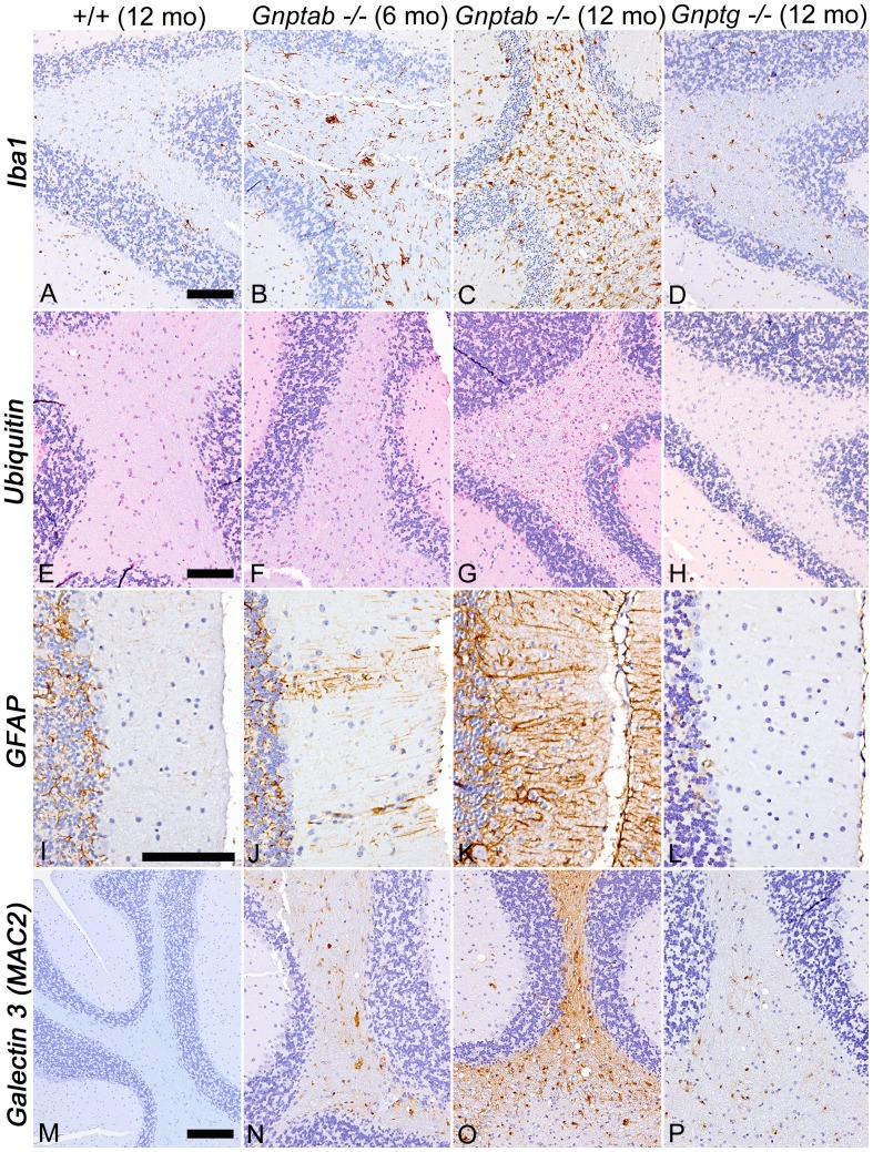
Figure 6. Cerebellar staining for ionized-calcium binding protein 1 (Iba1), ubiquitin (UBQ), glial fibrillary acidic protein (GFAP), and galectin-3 (MAC2) to detect microglia, ubiquinated proteins, astrocytes, and inflammation respectively.
(A-D), IHC staining for Iba1 showed small microglia with delicate cytoplasmic extensions in 12 month old WT tissue (A), diffuse reactive microgliosis In 6-month-old Gnptab−/− mice (B), markedly increased staining in 12-month-old Gnptab−/− mice (C), and only mild activation of microglia in 12-month-old Gnptg−/− mice (D). (E) Ubiquitin-positive granules were rarely detected in cerebellar white tracts of 12-month-old WT mice. (F,G) The amount and extent of ubiquitin-positive granules increased in severity between 6 and 12 months of age in Gnptab−/− mice. (H) There was a minimal increase in ubiquitin staining in 12-month-old Gnptg−/− mice. (I-L) IHC staining for GFAP showed faint staining of Bergmann glial cells in 12-month-old WT mice (I), mild multifocal activation of Bergmann glial cells in 6 month old Gnptab−/− mice (J), bilaterally symmetrical areas of Purkinje cell loss with associated thinning of the molecular layer and diffuse activation of Bergmann glia in 12-month-old Gnptab−/− mice (K) and no notable change in GFAP staining patterns involving Bergmann glia in 12-month-old Gnptg−/− mice (L). (M-P) IHC staining of galectin-3 (MAC2) was negative in the CNS tissues of WT mice (M), but showed progressive staining of cerebellar peduncles and white tracts of 6 and 12 month old Gnptab−/− mice (N,O). MAC2 reactivity was mild and mostly restricted to the white tracts and peduncles of the cerebellum in 12 month old Gnptg−/− mice (P). Figures A - H and M - P, 20X; Bar = 100 mm); Figures I - L, 40X; Bar = 100 mm).

