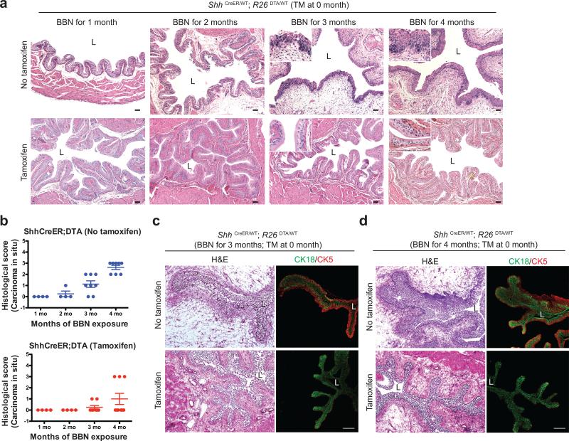
Figure 4. Ablation of Shh-expressing basal stem cells reduces CIS during nitrosamine-induced carcinogenesis while preserving urothelial architecture.
(a) Histopathological analysis (H&E) of TM-injected ShhCreER; R26DTA mice after 1, 2, 3, and 4 months of BBN exposure. Repeated experimental results are shown in Supplementary Table 2,3. (b) Histopathological evaluation of CIS in bladders from ShhCreER; R26DTA mice (Supplementary Table 2, 3). Scores from 0-3 are based on extent of CIS within urothelium (0, none; 1, <10%; 2, 10-30%; 3, >30%). No invasive carcinoma, and little or no CIS was induced by BBN exposure in animals with ablated stem cells. n=4 (1 month), n=4 (2 months), n=8 (3 months), n=8 (4months) for vehicle-treated ShhCreER; R26DTA mice; n=4 (1 month), n=4 (2 months), n=8 (3 months), n=9 (4months) for tamoxifen-treated ShhCreER; R26DTA mice. Data are presented as mean ± s.e.m (c,d) ShhCreER; R26DTA mice were injected with TM on five consecutive days to ablate Shh-expressing basal cells and exposed to BBN for 3 (c) and 4 (d) months. Sections from the bladders of control vehicle-injected or TM-injected mice (top and bottom panels, respectively) were stained for the luminal and basal markers, CK18 and CK5 (green and red, respectively). Note that lumenal cells are well-preserved at intermediate stages of BBN exposure in stem cell ablated animals. L, bladder lumen. Scale bars, 50μm.

