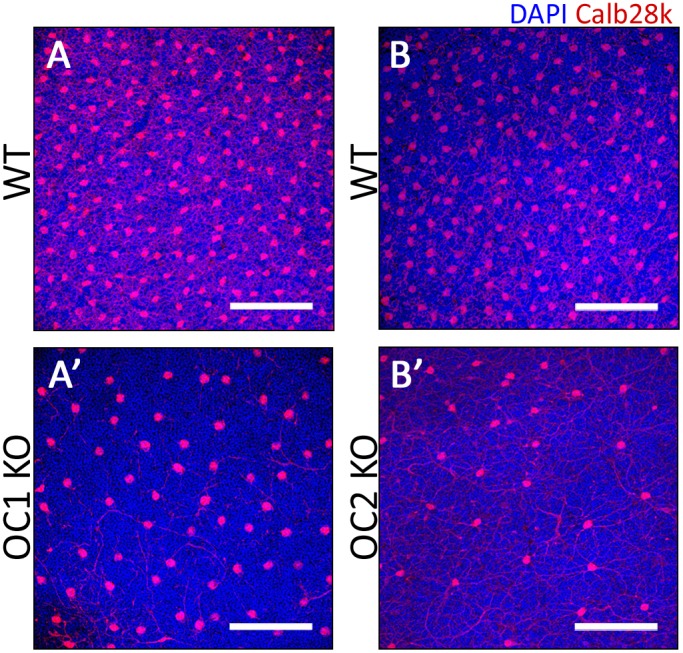
Figure 4. Assessment of horizontal cells by flat mount retina staining.

To better understand the distribution of horizontal cell loss in the OC1 and OC2-KO retinas, immunohistochemistry was performed using an anti-Calbindin 28k antibody on age-matched WT (A,B), OC1-KO (A’), and OC2-KO (B’) retinas. Scale bars represent 100 µm.
