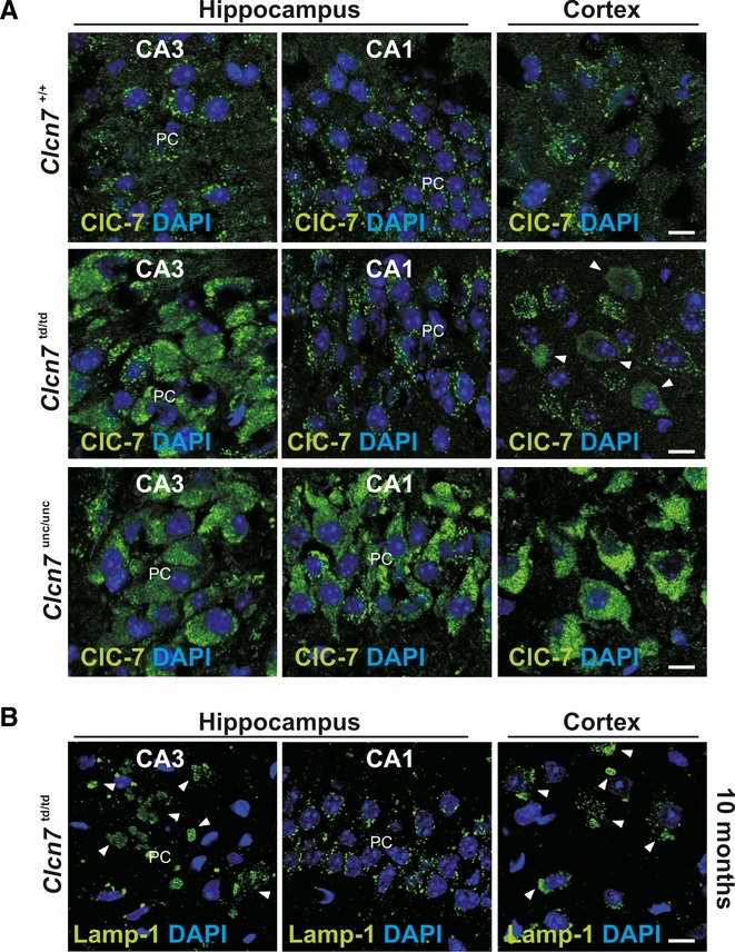
Figure 3. Abnormal lysosomal morphology in ClC-7 mouse models.

A ClC-7 immunolabelling in the somata of CA1 and CA3 pyramidal and of cortical neurons in Clcn7unc/unc, Clcn7td/td and WT mice. ClC-7td is abnormal in the CA3 region and partially in the cortex. Increased labelling intensity suggests larger ClC-7 amounts in Clcn7td/td CA3, and in Clcn7unc/unc CA3, CA1, and cortex (scale bar: 10 μm; PC, Purkinje cells).
B Abnormal Lamp-1 distribution in cortical and CA3 (arrowheads), but not CA1 neurons of 10-month-old Clcn7td/td mice. DNA stained with DAPI (scale bar: 10 μm).
