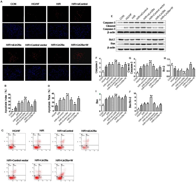
Figure 2. Antiapoptotic effect of Lin28a overexpression on cardiomyocytes after H/R injury in HG/HF conditions.
Representative images of TUNEL-stained primary neonatal cardiomyocytes after H/R stress in HG/HF conditions (A). Apoptosis of the primary cardiomyocytes determined by Cy3-annexinV/PI double staining and flow cytometry (n = 3). Region B2: late apoptotic cells (Cy3/PI, where Cy3 is cyanine-3 and PI is propidium iodide); Region B3: vital cells; Region B4: early apoptotic cells (C). Apoptotic index is expressed as the percentage of TUNEL-positive myocytes (in red) over total nuclei determined by DAPI staining(B). Apoptotic rate is expressed as the percentage of late apoptotic cells and early apoptotic cells(D). Protein expression with representative gel blots of Caspase-3, Cleaved Caspase-3, Bcl-2, Bax and β-actin (loading control) (E). Caspase-3(F); Cleaved Caspase-3(G); Bcl-2(H); Bax(I); Bax/Bcl-2 ratio(J). The columns and error bars represent means and SD. Data were obtained from at least three independent experiments.*P<0.05 vs. H/R, #P<0.05 vs. H/R+Lin28a.

