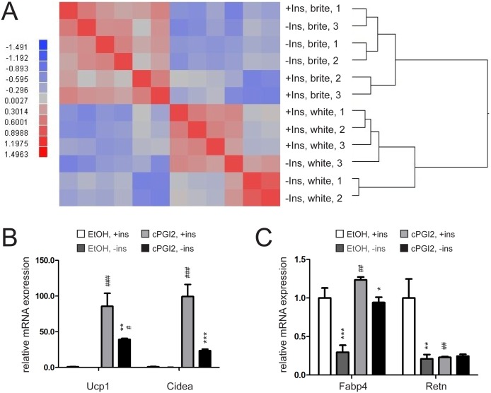
Figure 1. Lack of insulin impairs differentiation but not browning capacity of primary pre-adipocytes.
(A) Heatmap showing differential mRNA expression between confluent primary inguinal white adipose tissue (iWAT) precursor cells differentiated for 24 h with white (EtOH treated) or brite (cPGI2 treated) differentiation cocktail and between absence or presence of insulin (Ins) in the medium. Higher and lower expression is displayed in red and blue, respectively. (n = 3). (B) mRNA expression of UCP-1 and CIDEA or (C) FABP4 and RETN in primary iWAT precursor cells differentiated into white (EtOH treated) or brite (cPGI2 treated) adipocytes for 8 days with insulin present in the differentiation medium for the indicated timepoints (n = 3). All values in bar graphs are expressed as means ± SEM, #p<0.05, ##p<0.01, ###p<0.001 white (EtOH treated) vs. brite (cPGI2 treated) cells, *p<0.05, **p<0.01, ***p<0.001 normal conditions vs. insulin deprived conditions.

