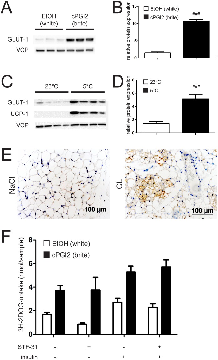
Figure 4. Browning induces GLUT-1 protein expression.
(A) Representative immunoblot and (B) imageJ quantification of GLUT-1 from primary inguinal white adipose tissue (iWAT) precursor cells. Cells were differentiated into white (EtOH treated) or brite (cPGI2 treated) adipocytes for 8 days. (C) Representative immunoblot and (D) imageJ quantification of GLUT-1 from primary inguinal white adipose tissue (iWAT) of mice housed at 23°C and 5°C respectively for 10 days. (E) GLUT-1 stained iWAT slices of mice fed LFD and implanted with NaCl or CL s.c. pumps. (F) 3H-2-deoxy-D-glucose (3H-2DOG) uptake by primary inguinal white adipose tissue (iWAT) precursor cells. Cells were differentiated into white (EtOH treated) or brite (cPGI2 treated) adipocytes for 8 days, ± GLUT-1 inhibitor STF-31 for the last 2 days and stimulated with 20 nM Insulin for 20 min. All values are expressed as means ± SEM, n = 7 for in vivo experiments and 10–12 animals for cell isolation.

