
Figure 4. Hrr25 kinase is responsible for Atg19 phosphorylation in the Atg11 binding region.
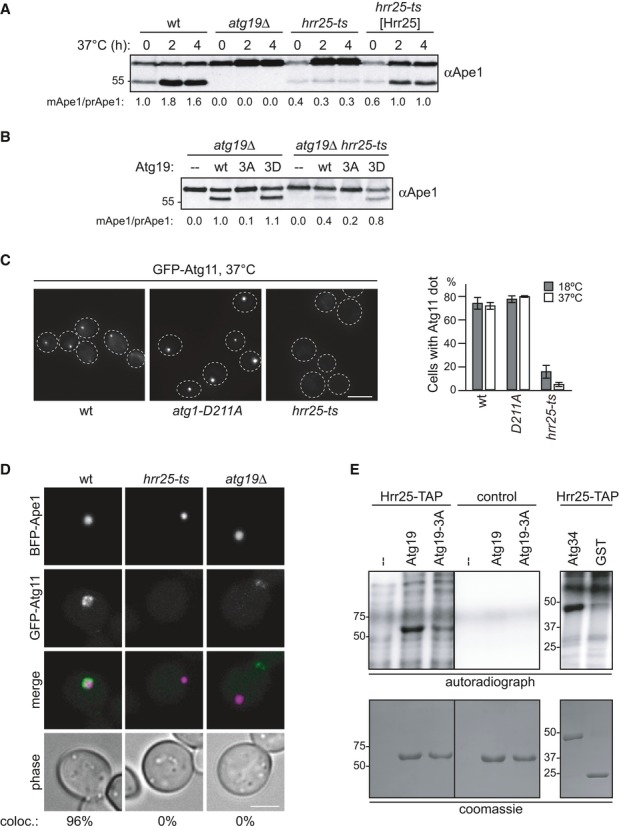
A Wild-type, hrr25-ts, and hrr25-ts cells containing HRR25 on a centromeric plasmid were grown to mid-log phase at 18°C followed by 2 and 4 h of log-phase growth at 37°C. Ape1 processing was analyzed by Western blotting.
B atg19Δ and atg19Δ hrr25-ts cells containing GFP-Atg19 as indicated were analyzed for Ape1 processing in log phase after 2 h at 37°C.
C GFP-Atg11, GFP-Atg11 hrr25-ts, and GFP-Atg11 atg1-D211A cells were analyzed for GFP-Atg11 dot formation. Quantification of at least 70 cells in three individual experiments is shown. Error bars represent standard deviation. Scale bar, 3 μm.
D GFP-Atg11, GFP-Atg11 hrr25-ts, and GFP-Atg11 atg19Δ cells containing BFP-Ape1 and Cup1-Ape1 were grown to log phase. Overexpression of Ape1 was induced by addition of 250 μM CuSO4 for 3 h. coloc.: Cells with GFP-Atg11/BFP-Ape1 co-localization. n = 50 (wt), 84 (hrr25-ts), 127 (atg19Δ). Scale bar, 3 μm.
E Hrr25 was immunoprecipitated from Hrr25-TAP cells and as a control from a non-tagged wild-type strain. In vitro phosphorylation of recombinant Atg19, Atg19-3A, Atg34, and GST using the kinase-bound beads was performed as described in Fig 3E. Both Atg19 panels are from the same gel with the same exposure.
Source data are available online for this figure.