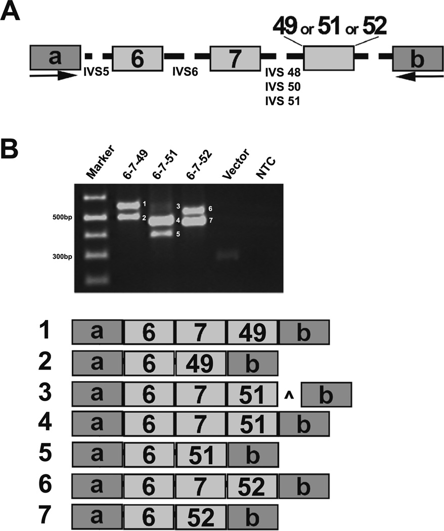
Fig. 5.
Evaluation of exon and intron signatures in Pkhd1. a For exon-trapping studies, minigene constructs containing mouse Pkhd1 exons 6, 7, and 49, 51, or 52 with 200 bp of flanking intronic sequences (shown as dashed lines) were cloned into the pSPL3 vector and transfected into COS7 cells. b Representative RT-PCR amplicons demonstrating the various splicing events is shown. Each reaction showed that exon 7 was skipped frequently (products 2, 5, and 7). In addition, there was a faint product 3, which included a 98-bp of PKHD1 IVS51 indicated by caret. a, b pSPL3 vector exons

