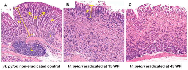
Figure 3. Representative gastric histopathology in H. pylori-infected p27 knockout mice.

A) A p27-deficient mouse infected with H. pylori SS1 strain for 70 weeks without receiving eradication treatment. There is an intense inflammatory cell infiltrate with lymphoid follicle formation and a replacement of the normal gastric glands by irregular dysplastic glands lacking the specialized chief and parietal cells. Arrows and D: dysplasia, height bar and H: hyperplasia (compared to normal foveolar length, “F” in panel B), I: inflammation, P: pseudopyloric metaplasia, L: lymphoid follicle.
B), C) p27-deficient mice that received H. pylori eradication therapy at 15 weeks (B) or 45 weeks (C) post infection. These mice have much less inflammation and retention of a normal gastric epithelial architecture, with no dysplasia. F: normal superficial foveolar epithelium.
Hematoxylin and eosin staining, original magnification × 100.
