Fig. 1.

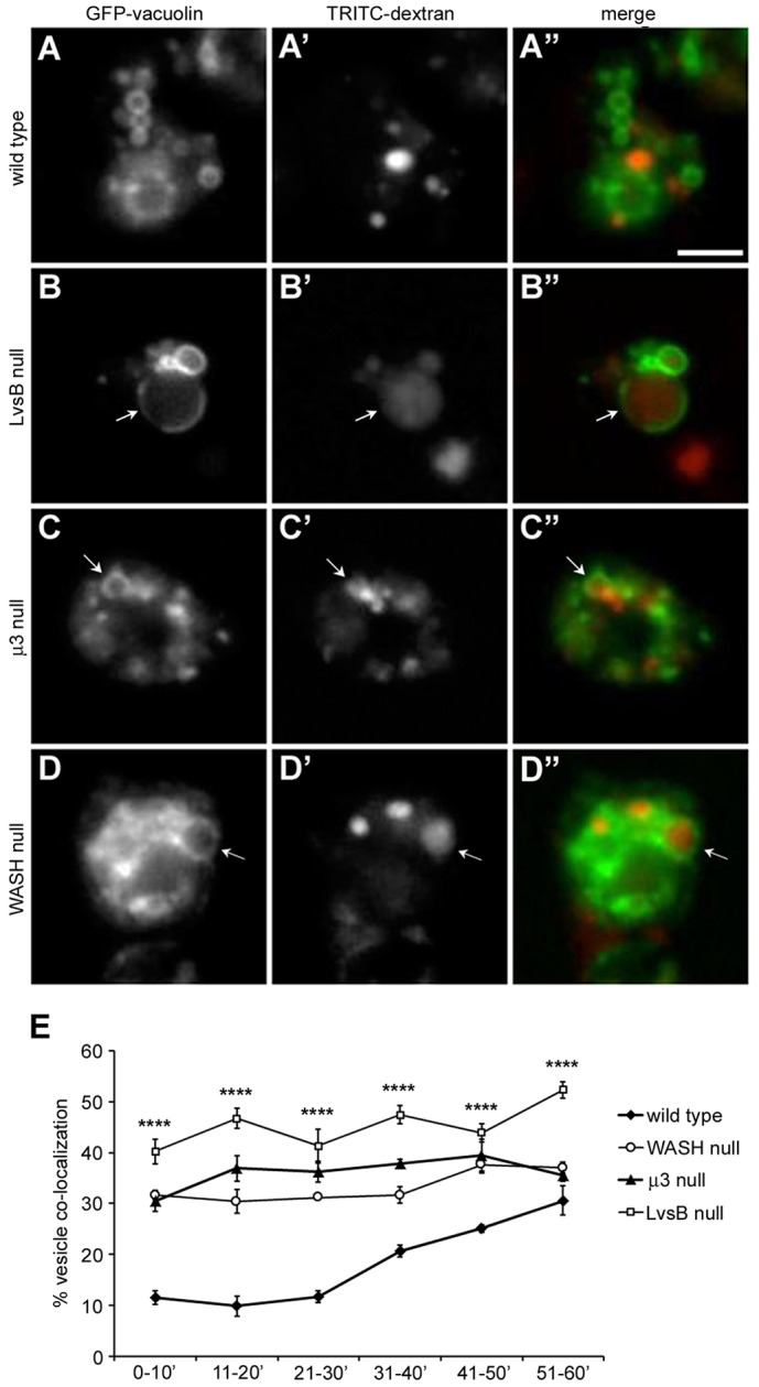
Vacuolin localizes to dextran-labeled vesicles at early time points in both LvsB-null and fission defect mutants. Cells were transfected with GFP–vacuolin and then given a 5-min pulse of TRITC–dextran to label a subpopulation of pinocytic vesicles. Cells were then washed and imaged continuously for 60 min. (A–D) Representative images selected from the 11–20-min time point are shown. (E) The percentage of GFP–vacuolin-labeled vesicles (>60 vesicles per experiment) containing TRITC–dextran was quantified for each 10-min interval and plotted as the mean±s.e.m. (n = 3). One-way ANOVA was used to calculate significant differences in the percentage of TRITC–dextran-labeled vesicles between cell lines for all time intervals shown. ****P<0.0001. During the first 30 min of imaging, the majority of wild-type cells maintained separation of their dextran and GFP–vacuolin-labeled vesicle populations (A–A″). In contrast, GFP–vacuolin was found to significantly overlap with TRITC–dextran at earlier time intervals in the LvsB-null (B–B″), μ3-null (C–C″) and WASH-null (D–D″) cell lines. Arrows indicate vesicles labeled by both TRITC–dextran and GFP–vacuolin. Scale bar: 5 µm.
