Fig. 2.
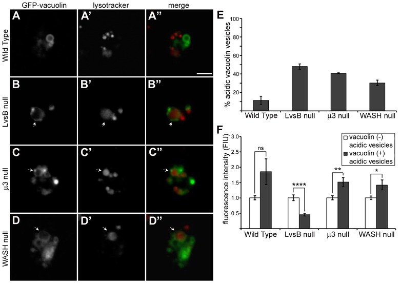
LvsB-null and fission defect mutants have an increased occurrence of acidic vacuolin-labeled vesicles but the characteristics of acidic vesicles in LvsB-null cells are distinct from those in fission defect mutants. (A–D) GFP–vacuolin-expressing cells were incubated with Lysotracker Red then imaged live to visualize acidic compartments with and without GFP–vacuolin membrane localization. (E) Using similar live-cell images, we quantified the percentage of GFP–vacuolin vesicles with Lysotracker Red fluorescence and plotted the mean±range for two experiments. As previously described, there was an increased occurrence of acidic GFP–vacuolin vesicles in LvsB-null cells (B–B″) compared to wild-type cells (A–A″). The fission defects in μ3-null cells (C–C″) and WASH-null cells (D–D″) also resulted in a higher percentage of acidic GFP–vacuolin vesicles similar to that observed in the LvsB-null cell line. (F) The fluorescence intensity of Lysotracker Red was measured in GFP–vacuolin-positive (late lysosomal or hybrid lysosomal organelles) and in GFP–vacuolin-negative (normal lysosomal) populations and used as an indicator of relative vesicle acidity. The fluorescence intensity of Lysotracker Red for each population was normalized within each cell line to the average fluorescence of GFP–vacuolin-negative lysosomes and plotted as the mean±s.e.m. ns, not significant, *P<0.05, **P<0.01, ****P<0.0001 (two-tailed Student's t-test). Notice that the relative acidity of vacuolin-labeled compared with unlabeled vesicles in LvsB-null cells is very different from that seen in wild-type, μ3-null, and WASH-null cells. The higher acidity of vacuolin-labeled vesicles in μ3-null and WASH-null cells is consistent with their role in vesicle fission during lysosomal maturation. In contrast, the fluorescence of GFP–vacuolin-positive vesicles is significantly reduced when compared to GFP–vacuolin-negative vesicles in the LvsB-null cells. This is consistent with a role for LvsB in reducing fusion between acidic lysosomal and neutral post-lysosomal compartments. Arrows indicate vesicles labeled by both Lysotracker Red and GFP–vacuolin. Scale bar: 5 µm.

