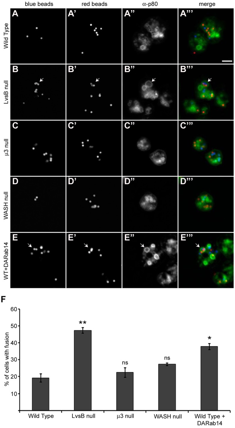
Fig. 5.

The phagosome defect of LvsB-null and DA-Rab14-expressing cells is not evident in the fission defect mutants. (A–E) Cells were given a pulse with blue fluorescent beads followed by a second pulse with red fluorescent beads to label adjacent populations of phagosomes. After a 30-min incubation to allow the labeled phagosomes to mature, cells were fixed, flattened and immunostained with anti-p80 antibody to label the limiting membranes of phagosomes. (F) The fraction of cells (>33 cells/experiment) with multi-particulate phagosomes containing both blue and red beads was plotted as the mean±s.e.m. of three experiments. A one-way ANOVA indicated significant differences in phagosome fusion between cell lines (P<0.0001). ns, not significant, *P<0.05, **P<0.01 (two-tailed Student's t-test with Bonferroni correction). The increased multi-particulate phagosome formation observed in both LvsB-null (B–B‴) and DA-Rab14-expressing (E–E‴) cells over wild-type cells (A–A‴) is consistent with previous studies and supports a fusion regulatory role for both proteins. The μ3-null (C–C‴) and WASH-null (D-D‴) fission defects did not induce significant increases in multi-particulate phagosome formation. This result suggests that a fission defect alone is not sufficient to cause the formation of multi-particulate phagosomes. Arrows indicate multi-particulate phagosomes. Scale bar: 5 µm.
