Fig. 8.
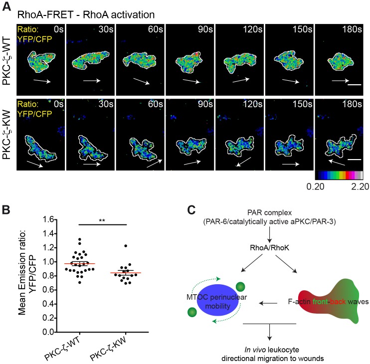
PKC-ζ regulates RhoA activity in leukocytes migrating to wounds in vivo. (A) Frames from representative movies of migrating myeloid cells in wounded tailfins are shown. The white arrows indicate the direction of migration. Cell outlines are shown as white lines. Scale bars: 10 µm. See also supplementary material Movie 12. RhoA activity was visualized in TG(FmpoP::mCherry) larvae transiently expressing cytosolic RhoA-FRET biosensor together with PKC-ζ-WT or PKC-ζ-KW in myeloid cells. Ratiometric images of YFP∶CFP emission for each cell are shown. (B) Average activation level of RhoA (mean emission ratio of YFP∶CFP for the entire cell during migration) in wound-activated leukocytes. Data are expressed as the mean±s.e.m. of all analyzed cells (PKC-ζ-WT, 25 leukocytes in 15 larvae; PKC-ζ-KW, 15 leukocytes in 5 larvae); **P<0.01 (two-tailed unpaired Student's t-test). (C) A model illustrating the mechanism by which the PAR complex controls wound-directed leukocyte migration in vivo. The PAR complex coordinately controls Rho-dependent F-actin dynamics and MTOC perinuclear mobility to support the persistent migration of leukocytes to wounds.

