Fig. 2.
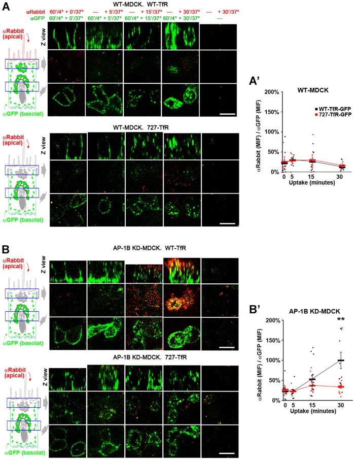
The N-glycan linked to N727 mediates apical transcytosis of TfR in AP-1B KD MDCK cells. Polarized WT (A) and AP-1B KD (B) MDCK cells were transiently transfected with either WT (top) or N727A (bottom) TfR–GFP. SeTau647-labeled rabbit anti-GFP antibodies (αGFP, green) were applied to the basolateral chamber (120 min, 4°C) to allow basolateral surface binding. The temperature was shifted to 37°C to allow antibody uptake and transcytosis for the indicated times periods, during which time the Alexa-Fluor-568-conjugated anti-rabbit-IgG antibody (αRabbit, red) was applied to the apical chamber. (A′,B′) Cells from experiments represented in A and B were quantified for the ‘αRabbit (MIF):αGFP (MIF) ratio, which is proportional to the fraction of basolateral SeTau647-labeled rabbit anti-GFP antibody transcytosed to the apical plasma membrane. Values were normalized to the highest value (i.e. 30 min uptake in AP-1B KD MDCK with WT-TfR–GFP). Circles correspond to individual cells obtained from different experiments and thick lines indicate the mean±s.e.m. **P<0.001. Scale bars: 10 µm.

