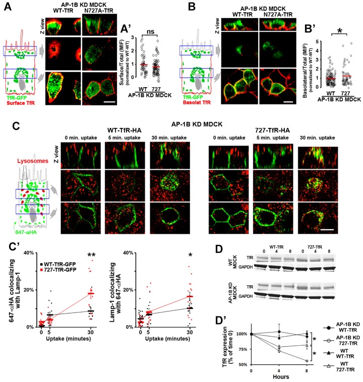
Fig. 4.
N727A TfR recycles inefficiently to the basolateral membrane and is targeted for lysosomal degradation in AP-1B KD MDCK cells. (A) Polarized AP-1B KD MDCK cells were transiently transfected with either WT- or N727A-TfR–GFP and were immunostained for surface TfR–GFP (without permeabilizing the cells), with primary rabbit anti-GFP (green) and secondary Alexa-Fluor-568-conjugated anti-rabbit-IgG (red) antibodies applied to both the apical and basolateral chambers. (A′) Cells from experiments represented in A were quantified for the surface:total ratio using the fluorescent signals of Alexa-Fluor-568-conjugated anti-rabbit-IgG (surface) and GFP (total). (B) Polarized AP-1B KD MDCK cells were transiently transfected with either WT- or N727A-TfR–GFP and immunostained for basolateral TfR–GFP only. (B′) Cells from experiments represented in B were quantified for the basolateral:total ratio using the fluorescent signals of Alexa-Fluor-568-conjugated anti-rabbit-IgG (basolateral) and GFP (total). (C) Polarized AP-1B KD MDCK cells were transiently transfected with either WT- or N727A-TfR–HA. SeTau647-labeled anti-HA antibody (647-αHA, green) was bound to the basolateral plasma membrane (120 min, 4°C), the temperature was shifted to 37°C to allow antibody uptake and cells were fixed at the indicated times periods and immunostained for the lysosomal marker Lamp1. (C′) Cells from experiments represented in C were quantified for the percentage of pixels of 647-αHA colocalizing with Lamp1 (left) and the percentage of pixels of Lamp1 colocalizing with 647-αHA (right), for each time point studied. (D) WT and AP-1B KD MDCK cells were nucleofected with either WT- or N727A-TfR–GFP, treated with cycloheximide for the indicated time periods and analyzed for TfR–GFP expression by western blotting. (D′) Quantitative analysis of WT- and N727A-TfR–GFP in three or four experiments represented in D. Circles correspond to individual cells obtained from different experiments and thick lines indicate the mean±s.e.m. for C′ and D′. ns, not significant; * P<0.05; ** P<0.01. Scale bars: 10 µm.

