Fig. 5.
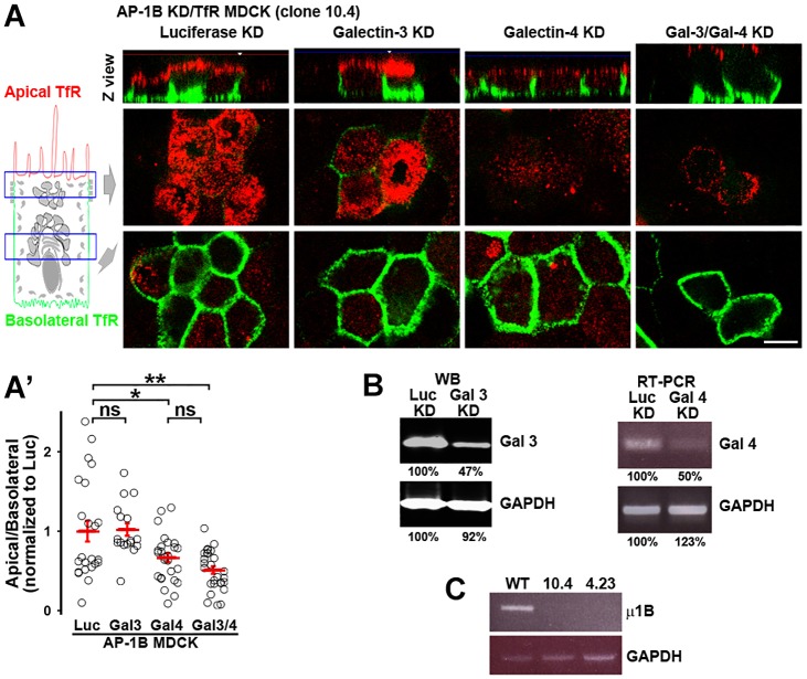
Galectin-4 mediates apical localization of TfR in AP-1B KD MDCK cells. (A) AP-1B KD MDCK cells stably expressing human TfR (AP-1B KD/TfR MDCK) were knocked down for luciferase (Luc), galectin-3 (Gal 3), galectin-4 (Gal 4), or both galectin-3 and galectin-4 (Gal3/4) and polarized on transwell filters. Apical and basolateral TfR was immunostained (without permeabilizing the cells), using anti-human-TfR primary antibody that recognizes the luminal domain of TfR and Alexa-Fluor-568-conjugated anti-mouse-IgG (red) and Alexa-Fluor-647-conjugated anti-mouse-IgG (green) secondary antibodies in the apical and basolateral chambers, respectively. (A′) Cells from experiments represented in A were quantified for the apical:basolateral ratio using the fluorescence signal of each plasma membrane domain. (B) Western blot (WB) analysis of galectin-3 expression, and RT-PCR analysis of galectin-4 expression in AP-1B KD/TfR MDCK cells electroporated with siRNA against luciferase, galectin-3 or galectin-4. A quantification of protein levels is presented below the blot. (C) RT-PCR analysis of μ1B expression (the medium subunit of AP-1B) in WT MDCK and the two AP-1B KD/TfR MDCK clones used in this work. Circles correspond to individual cells obtained from different experiments and red lines indicate the mean±s.e.m. ns, not significant; *P<0.05; **P<0.001. Scale bar: 10 µm.

